Volume: 1 | Issue: 1 | View as PDF
  • OPEN ACCESS

The Physiological and Pathological Roles of D-serine in the Retina

  • Pianshi Zhou1,2 and
  • Shengzhou Wu1,2,* 
 Author information
Nature Cell and Science   2023;1(1):9-15

doi: 10.61474/ncs.2023.00003

Abstract

D-serine (D-ser) is highly enriched in the central nervous system and is a neuromodulator that acts on glutamate-bound N-methyl-D-aspartate receptor (NMDAR), playing vital roles in brain physiology and neuropsychiatric disorders. The levels of D-Ser are principally determined by serine racemase (SR) and D-amino acid oxidase (DAAO), and the regulations by these enzymes show regional differences in the brain. Reduction of D-Ser by chemicals or genetic modulation of SR or DAAO greatly attenuates NMDAR activity and the ensued excitotoxicity, a shared mechanism in neurodegenerative disorders and stroke. In the inner retina, D-Ser inhibits non-NMDA glutamate receptors and recruits NMDAR, thus potentiating the NMDAR-dependent current in retinal ganglion cells (RGCs). In the outer retina, D-Ser participates in vertical signal processing and maintains the retinal functional integrity. Accumulated studies suggest that D-Ser plays a role in diabetic retinopathy and glaucoma; in both disorders, RGC demise is a shared pathology. Loss of SR alleviates RGC death and retinal microvascular abnormalities in diabetic rodents, and increasing DAAO blunts RGC death in glaucoma animals. Thus, regulation of D-Ser availability by chemicals or genetic modulation of SR or DAAO may serve as a promising strategy to treat ocular disorders involving abnormal activation of NMDAR. This mini-review begins with the roles of D-Ser in the brain, extends the topic to the retina, and finally discusses relevant research progress in diabetic retinopathy and glaucoma. This is the first review highlighting the role of the NMDAR co-agonist D-Ser in the physiopathology of the retina.

Keywords

D-serine, Serine racemase, NMDAR, D-amino acid oxidase, Diabetic retinopathy, Excitotoxicity, Glaucoma

Introduction

D-serine (D-ser) functions as a co-agonist of the glutamate-bound N-methyl-D-aspartate receptor (NMDAR), regulating neurotransmission and synaptic plasticity. Through this channel-type receptor, D-Ser modulates long-term potentiation, neuronal migration, and excitotoxicity.1–6 To potentiate NMDAR activity, D-Ser binds to the GluN1 subunit, in concert with GluN2-bound glutamate, opening the receptor and allowing the influx of sodium or calcium.1,4

D-Ser in the brain

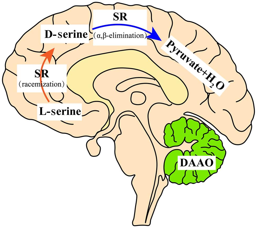
D-Ser is primarily generated from levorotary serine (L-Ser) through a racemization reaction by serine racemase (SR), a pyridoxal 5′-phosphate-dependent enzyme, and degraded through oxidative deamination by the peroxisomal enzyme D-amino acid oxidase (DAAO), a flavine adenine dinucleotide-containing enzyme.1,7 SR also degrades D-Ser by α,β-elimination dehydration.8 In rodents and humans, SR is enriched in the forebrain while DAAO is substantially expressed in the hindbrain and spinal cord.9–11 Correspondingly, D-Ser is enriched in the forebrain and present in lesser amounts in the hindbrain and spinal cord in adults.11 Regulation of D-Ser by SR and DAAO shows regional differences in the brain because of the distributional difference between the two enzymes. In the rodent forebrain, knockout of SR reduces the D-Ser level by 90%, whereas loss of DAAO does not change the D-Ser level in this area.12 On the other hand, loss of DAAO increases the D-Ser level by more than 20 times in the hindbrain and spinal cord, while knockout of SR minimally changes the D-Ser level in these areas.12,13 Thus, SR determines the D-Ser level in the forebrain, and DAAO controls the level in the hindbrain and spinal cord.

Tracing the cellular origin of D-Ser, SR is mostly confined to protoplasmic astrocytes according to the early studies; however, recent data indicate that SR is mostly localized to glutamatergic neurons, although reactive astrocytes and microglia express some SR.6,14–19 Regarding the cellular distribution, DAAO is preferentially confined to astroglia in the hindbrain.20 The distribution of D-Ser in the central nervous system is depicted in Figure 1.

Distribution and metabolism of D-serine in the mammalian brain.
Fig. 1  Distribution and metabolism of D-serine in the mammalian brain.

In the forebrain, D-serine is abundant. SR synthesizes D-serine via racemization, while D-serine is degraded via an α,β-elimination reaction because of DAAO deficiency in the forebrain. In the adult cerebellum, D-serine is less abundant due to DAAO degradation. DAAO, D-amino acid oxidase; SR, serine racemase.

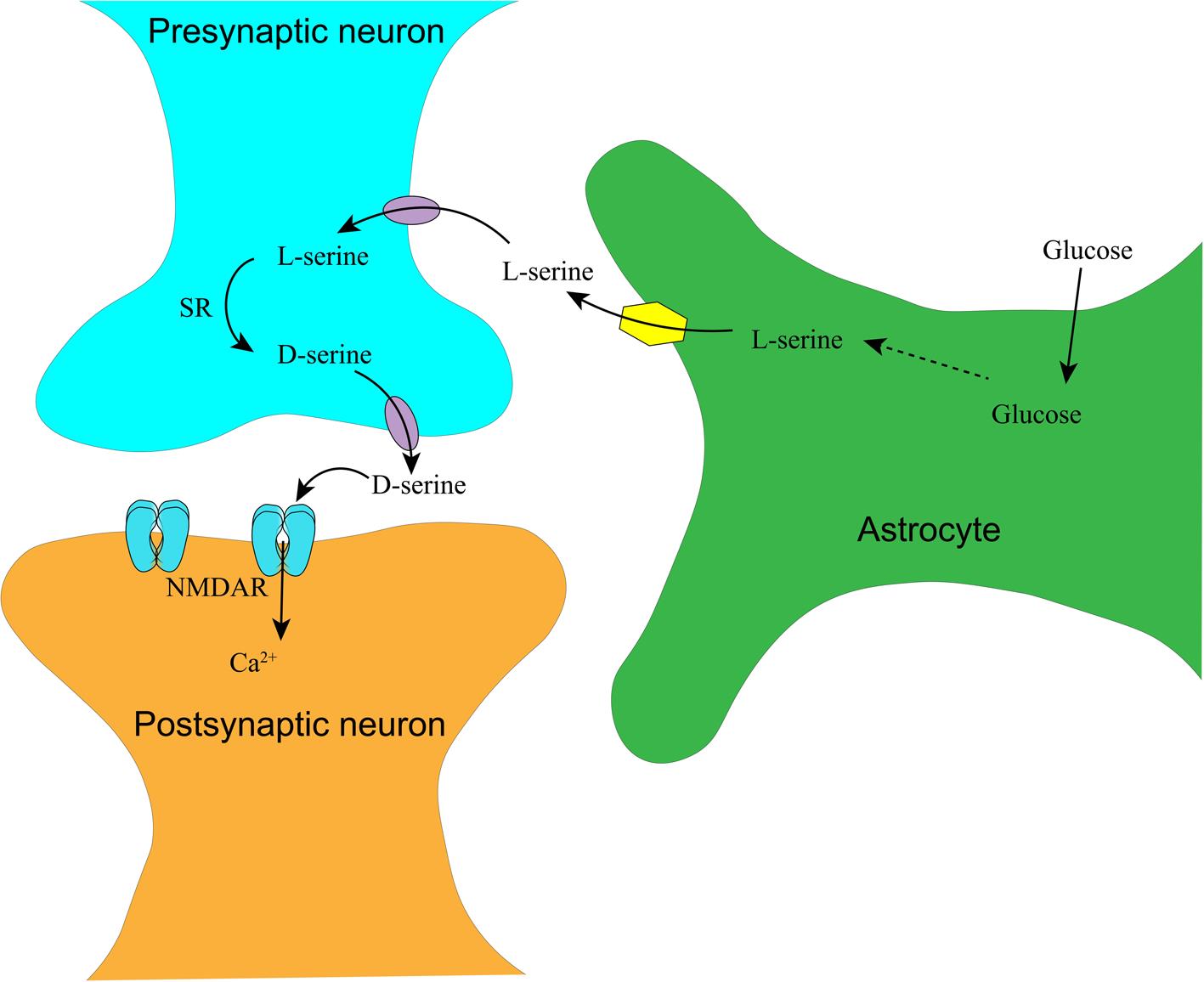
Appropriate NMDAR activity is also dependent on serine transport between astrocytes and neurons. To explain this transport, Herman Wolosker has proposed a delicate model dissecting serine transport in the brain: astrocytes take up glucose from the peripheral blood and metabolize it to L-Ser. L-Ser is then transported into presynaptic neurons, where it is converted to D-Ser by SR. D-Ser is transported out and acts on the NMDAR on the postsynaptic membrane.21 The synthesis and transportation of D-Ser in the central nervous system are depicted in Figure 2.

Synthesis and transport of D-serine in the central nervous system.
Fig. 2  Synthesis and transport of D-serine in the central nervous system.

In the tripartite synapse consisting of pre-/post-synaptic neurons and enveloping astrocytes, glucose is taken up into astrocytes via glucose transporter 1 and synthesized into L-serine. L-serine is transported into the presynaptic neuron via a neural amino acid transporter, where L-serine is converted into D-serine by SR. Released D-serine and glutamate from the presynaptic neuron act on NMDAR on the postsynapse, triggering calcium or sodium influx. NMDAR, N-methyl-D-aspartate receptor; SR, serine racemase.

Inappropriate activity of NMDAR is implicated in neuropsychiatric disorders, including Alzheimer’s disease and schizophrenia.22–24 Overactivation of NMDAR leads to excitotoxicity, a shared mechanism in neurodegenerative disorders and stroke; whereas hypofunction of NMDAR is associated with schizophrenia.23–25 The use of high-affinity and strong competitive antagonists of NMDAR targeting the GluN2 subunit leads to gross neurological impairment and behavioral toxicity, whereas the side effects of uncompetitive antagonists are moderate.26 Thus, regulation of D-Ser availability seems much more tolerable. Blockade of D-Ser on the glycine-binding site of NMDAR or depletion of D-Ser by chemicals or genetic manipulation effectively attenuates NMDAR activity and greatly reduces excitotoxicity.4,6,27,28 For example, knockout of SR substantially protects against cerebral ischemia in rodents and, in doing so, greatly reduces injury in the NMDA/amyloid peptide-injected brain in rodents.28,29 Similarly, a mutation in DAAO leading to excessive production of D-Ser is associated with motor neuron degeneration in amyotrophic lateral sclerosis.30

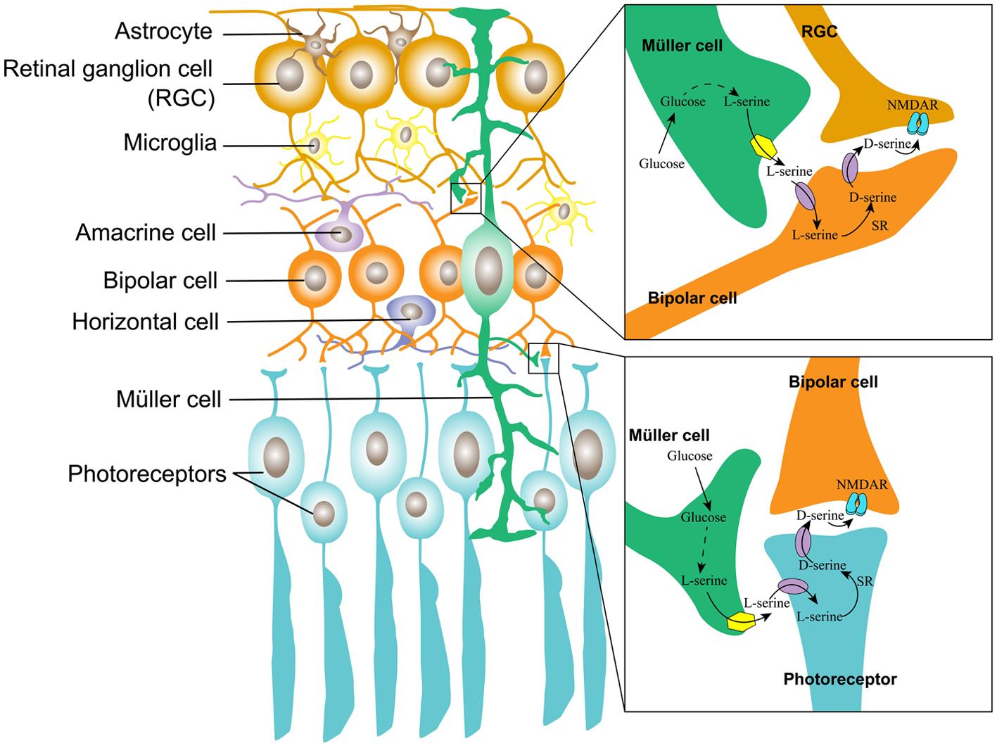
The Müller cell is the major type of glial cell in the retina and the outer retina; it provides trophic and anti-oxidative support for photoreceptors as well as maintains the inner blood-retinal barrier. In the inner retina, Müller cells regulate synaptic activity by uptaking glutamate.31 By mimicking the serine shuttle between astrocytes and neurons in the brain, we propose a similar model in the retina, which is depicted in Figure 3.

The serine shuttle in the retina.
Fig. 3  The serine shuttle in the retina.

The Müller cell constitutes a tripartite synapse in the inner and outer retina. The Müller cell takes up glucose from the blood and metabolizes glucose to L-serine. In the inner retina, L-serine is transported into a bipolar cell, where it is converted into D-serine, which is transported out and acts upon NMDAR on the RGC; in the outer retina, L-serine is transported into the photoreceptor, where it is converted by SR into D-serine, which is released to the synaptic cleft, acting upon NMDAR on the bipolar cells. NMDAR, N-methyl-D-aspartate receptor; RGC, retinal ganglion cell.

The physiological roles of D-Ser in the retina

In the retina, D-Ser and SR are expressed in astroglia, Müller glia, retinal ganglion cells, and retinal pigment epithelial (RPE) cells.32–34 By comparison, SR in the retina is expressed at approximately the same level as that in the cortex.32 In detail, SR expression is greater in retinal neurons than in RPE cells.35 Early studies indicate that DAAO activity is at a relatively higher level in the inner plexiform layer of the rodent retina and also in the peroxisome of RPE cells and Müller glia in the amphibian retina.36,37 Our recent study also indicates that DAAO is mostly expressed in the RGCs, outer plexiform layer, and RPE cells in the rodent retina.38 In addition, knockout of SR decreases the D-Ser level in the mature retina by 90% relative to that of wild-type mice; while in the DAAO mutant line, the D-Ser level in the retina increased by only two-fold compared with that of wild-type mice.39 These analyses suggest that in the rodent retina, similar to the brain, D-Ser is principally synthesized by SR; while DAAO is not the only enzyme to degrade D-Ser in the retina, different from the dominant effect of DAAO on D-Ser degradation in the hindbrain and spinal cord.12

The function of D-Ser in the retina is much less clear than in the brain. In the mammalian retina, functional NMDAR is mostly localized to RGCs, the inner nuclear layer, and photoreceptors.32,40 In the inner retina, D-Ser inhibits non-NMDA glutamate receptors, recruits NMDAR, and mediates light-induced RGC current.36–41 Deletion of SR removes the NMDA component of the light-induced synaptic response but does not affect visual function.42 Meanwhile, in DAAO-knockout mice, exogenous application of D-Ser potentiates the NMDAR current in RGCs, suggesting that DAAO mediates subsaturation of the NMDAR glycine site in the retina.36 In contrast, the functional roles of D-Ser in the outer retina are less clarified. NMDAR hypo- and hyperfunction in the retina alter the components of the flash electroretinogram. The use of this technique demonstrated that deletion of SR significantly delayed the a-wave and reduced the b-wave amplitude, and the effects were more prominent in male Srr-knockout mice; meanwhile, the application of exogenous D-Ser through overactivation of NMDAR similarly alters the retinal field potential, but these changes are not observed in female wild-type mice.43 These differences suggest that the glycine-binding site of NMDAR is less saturated in the retina of male mice.

D-Ser may play a role in the early development of the retina. The neonatal retina consists of a double-cell layer, which is composed of an inner and an outer neuroblastic layer, that is separated by the inner plexiform layer, and it grows into a triple-cell layer at two weeks after birth, with a structure similar to that of the adult retina.44 D-Ser in the retina peaks in neonatal mice, followed by a dramatic decrease as the mice grow into adults, suggesting that D-Ser may play a role in the early development of the retina.33,39

The pathogenic roles of D-Ser in diabetic retinopathy (DR) and glaucoma

DR affects one-third of diabetic patients when the duration of diabetes lasts over a decade.45 The characteristics of DR include the loss of retinal vascular endothelial cells and pericytes as well as microaneurysm formation in the early stage and preretinal/vitreous hemorrhage, retinal neovascularization, and retinal detachment at the later stage of DR.46 It has taken a long time to recognize that DR is also a neurodegenerative disorder, and the neurodegeneration in the retina antedates overt retinal microvascular pathologies in diabetic animals and humans.47–50 The pathologic cascade of DR possibly begins with a retinal neurovascular uncoupling, retinal neurodegeneration, and reactive gliosis, followed by the ensuing disruption of the blood-retinal barrier and more severe retinal microvascular abnormalities.51,52 Conversely, disruption of the blood-retinal barrier causes infiltration of blood-born leukocytes and activation of residential microglia, which can compromise neuronal function and viability via cytokines and oxidative stress.53,54 Thus, neurodegeneration and injuries to the retinal microvessels mutually aggravate during DR progression, culminating in avalanche-like abnormalities at the end stage of DR.

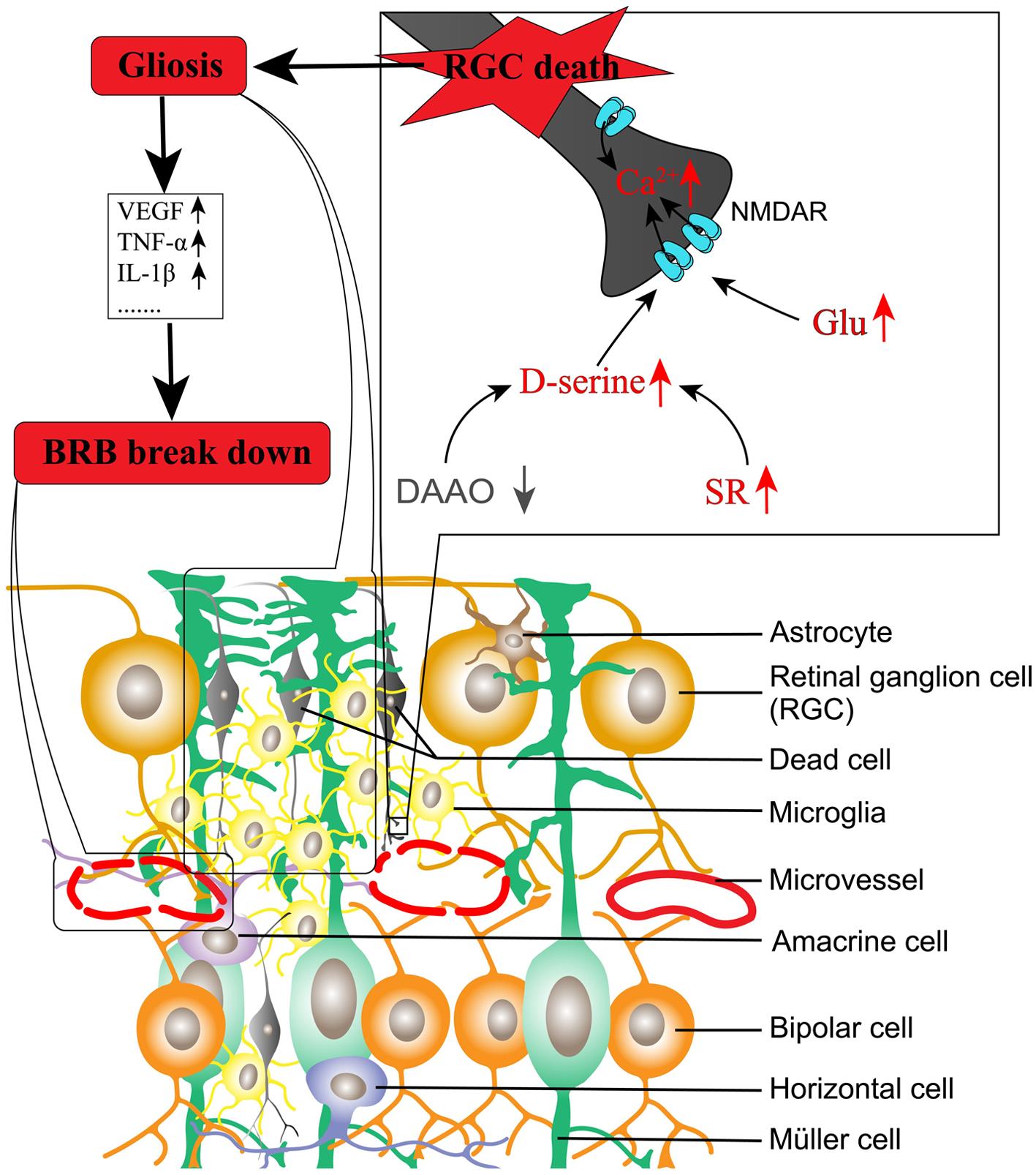
NMDAR activation is intimately associated with retinal neurodegeneration. In diabetes, the activity of glutamate transporter expressed on Müller glia is significantly decreased by oxidative stress and thus impairs glutamate metabolism.55,56 Diabetes also increases the expression of ionotropic glutamate receptor subunits in the human and rat retina.57,58 Meanwhile, an uncompetitive antagonist of NMDAR used for treating Alzheimer’s disease has been demonstrated to protect against retinal neurodegeneration and microvascular damage in diabetic rats.59 Considering that depletion of D-Ser largely attenuates NMDAR-mediated calcium influx and excitotoxicity,4,6 we explored the roles of SR and D-Ser in DR. Our results indicated the following: (1) SR is increased in the diabetic rat retina;60 (2) D-Ser is increased in the aqueous humor in diabetic rats and the aqueous and vitreous humor in diabetic humans;60,61 (3) loss of SR significantly reduces retinal neurodegeneration mediated by injected NMDA and in Ins2Akita mice, an animal model of type 1 diabetes;62,63 and (4) DAAO is decreased in the retina of diabetic rats, and overexpression of DAAO in the retina delivered by adeno-associated virus 8 prevents retinal neurovascular changes in diabetic rats.38 A study from another group also indicates that knockout of SR significantly reduces retinal neurodegeneration and loss of endothelial cells in retinal microvessels in diabetic mice.64 We propose that the imbalanced production of D-Ser coupled with extracellular glutamate in the diabetic retina contributes to DR. The model is depicted in Figure 4.

Imbalanced production of D-serine contributes to diabetic retinopathy.
Fig. 4  Imbalanced production of D-serine contributes to diabetic retinopathy.

In models of diabetic retinopathy, SR is increased while DAAO is decreased in the retina compared to normal animals, leading to the increased production of D-serine. D-serine, coupled with increased glutamate in the extracellular compartment due to impaired glutamate transport, activates NMDAR, leading to RGC death. Consequently, glia including Müller cells, astrocytes, and microglial cells proliferate and release pro-inflammatory factors and pro-angiogenic factors, resulting in disruption of the blood-retinal barrier. BRB, blood-retinal barrier; DAAO, D-amino acid oxidase; Glu, glutamate; IL-1, interleukin-1; NMDAR, N-methyl-D-aspartate receptor; RGC, retinal ganglion cell; SR, serine racemase; TNF, tumor necrosis factor; VEGF, vascular endothelial growth factor.

Similar to DR, glaucoma is a leading ocular disorder resulting in blindness worldwide. Long-term intraocular hypertension or the imbalance of the trans-lamina cribrosa pressure is associated with RGC degeneration and RGC axonopathy in glaucoma.65,66 Recently, studies from the same group have reported that D-Ser is significantly increased in the retina of glaucomatous animals and that depletion of D-Ser by DAAO prevents apoptosis of RGCs.67,68

Conclusion

Accumulated studies suggest that D-Ser is involved in the pathogenesis of DR and glaucoma. Therefore, regulation of D-Ser availability may provide a promising strategy to prevent or treat these disorders.

Abbreviations

BRB: 

blood-retinal barrier

DAAO: 

D-amino acid oxidase

DR: 

diabetic retinopathy

D-Ser: 

D-serine

Glu: 

glutamate

IL-1: 

interleukin-1

L-Ser: 

L-serine

NMDAR: 

N-methyl-D-aspartate receptor

RGC: 

retinal ganglion cell

RPE: 

retinal pigment epithelial

SR: 

serine racemase

TNF: 

tumor necrosis factor

VEGF: 

vascular endothelial growth factor

ATP: 

adenosine triphosphate

CVD: 

cardiovascular disease

CM: 

cardiomyopathy

IS: 

ischemic stroke

mtDNA-CN: 

mtDNA copy number

mtDNA: 

mitochondrial DNA

MELAS: 

mitochondrial neuro-gastro-intestinal encephalomyopathy

MUTYH: 

mutY DNA glycosylase

nDNA: 

nuclear DNA

OGG1: 

8-oxoguanine DNA glycosylase

OS: 

oxidative stress

OXPHOS: 

oxidative phosphorylation system

POLG: 

polymerase subunit gamma

ROS: 

reactive oxygen species

TFAM: 

mitochondrial transcription factor A

8oxoG: 

7,8-dihydro-8-oxoguanine

Declarations

Acknowledgement

None.

Funding

The related studies have been supported by start-up funding (89210001) from Wenzhou Medical College, the National Natural Science Foundation of China (81371027), and an intramural grant: Integrated Project of the State Key Laboratory of the School of Optometry and Ophthalmology (#J02-20190204) to Dr. Shengzhou Wu.

Conflict of interest

The manuscript was submitted during Dr. Shengzhou Wu’s term from May 2023 to December 2024 serving as an editorial board member of Nature Cell and Science. The authors have no other conflicts of interest to declare.

Authors’ contributions

Critical revision of the manuscript for important intellectual content, administrative, technical, or material support (ZP). Study concept and design, drafting of the manuscript (WS).

References

  1. Wolosker H, Blackshaw S, Snyder SH. Serine racemase: a glial enzyme synthesizing D-serine to regulate glutamate-N-methyl-D-aspartate neurotransmission. Proc Natl Acad Sci U S A 1999;96(23):13409-13414 View Article PubMed/NCBI
  2. Panatier A, Theodosis DT, Mothet JP, Touquet B, Pollegioni L, Poulain DA, et al. Glia-derived D-serine controls NMDA receptor activity and synaptic memory. Cell 2006;125(4):775-784 View Article PubMed/NCBI
  3. Turpin FR, Potier B, Dulong JR, Sinet PM, Alliot J, Oliet SH, et al. Reduced serine racemase expression contributes to age-related deficits in hippocampal cognitive function. Neurobiol Aging 2011;32(8):1495-1504 View Article PubMed/NCBI
  4. Mothet JP, Parent AT, Wolosker H, Brady RO, Linden DJ, Ferris CD, et al. D-serine is an endogenous ligand for the glycine site of the N-methyl-D-aspartate receptor. Proc Natl Acad Sci U S A 2000;97(9):4926-4931 View Article PubMed/NCBI
  5. Zhang H, Song L, Chang Y, Wu M, Kuang X, Jiang H, et al. Potential deficit from decreased cerebellar granule cell migration in serine racemase-deficient mice is reversed by increased expression of GluN2B and elevated levels of NMDAR agonists. Mol Cell Neurosci 2017;85:119-126 View Article PubMed/NCBI
  6. Wu SZ, Bodles AM, Porter MM, Griffin WS, Basile AS, Barger SW. Induction of serine racemase expression and D-serine release from microglia by amyloid beta-peptide. J Neuroinflammation 2004;1(1):2 View Article PubMed/NCBI
  7. Sasabe J, Miyoshi Y, Suzuki M, Mita M, Konno R, Matsuoka M, et al. D-amino acid oxidase controls motoneuron degeneration through D-serine. Proc Natl Acad Sci U S A 2012;109(2):627-632 View Article PubMed/NCBI
  8. Foltyn VN, Bendikov I, De Miranda J, Panizzutti R, Dumin E, Shleper M, et al. Serine racemase modulates intracellular D-serine levels through an alpha,beta-elimination activity. J Biol Chem 2005;280(3):1754-1763 View Article PubMed/NCBI
  9. Horiike K, Tojo H, Arai R, Nozaki M, Maeda T. D-amino-acid oxidase is confined to the lower brain stem and cerebellum in rat brain: regional differentiation of astrocytes. Brain Res 1994;652(2):297-303 View Article PubMed/NCBI
  10. Miyoshi Y, Hamase K, Okamura T, Konno R, Kasai N, Tojo Y, et al. Simultaneous two-dimensional HPLC determination of free D-serine and D-alanine in the brain and periphery of mutant rats lacking D-amino-acid oxidase. J Chromatogr B Analyt Technol Biomed Life Sci 2011;879(29):3184-3189 View Article PubMed/NCBI
  11. Suzuki M, Imanishi N, Mita M, Hamase K, Aiso S, Sasabe J. Heterogeneity of D-Serine Distribution in the Human Central Nervous System. ASN Neuro 2017;9(3):1759091417713905 View Article PubMed/NCBI
  12. Miyoshi Y, Konno R, Sasabe J, Ueno K, Tojo Y, Mita M, et al. Alteration of intrinsic amounts of D-serine in the mice lacking serine racemase and D-amino acid oxidase. Amino Acids 2012;43(5):1919-1931 View Article PubMed/NCBI
  13. Hashimoto A, Nishikawa T, Konno R, Niwa A, Yasumura Y, Oka T, et al. Free D-serine, D-aspartate and D-alanine in central nervous system and serum in mutant mice lacking D-amino acid oxidase. Neurosci Lett 1993;152(1-2):33-36 View Article PubMed/NCBI
  14. Schell MJ, Molliver ME, Snyder SH. D-serine, an endogenous synaptic modulator: localization to astrocytes and glutamate-stimulated release. Proc Natl Acad Sci U S A 1995;92(9):3948-3952 View Article PubMed/NCBI
  15. Beltrán-Castillo S, Triviño JJ, Eugenín J, von Bernhardi R. TGFβ1-Smad3 signaling mediates the formation of a stable serine racemase dimer in microglia. Biochim Biophys Acta Proteins Proteom 2020;1868(9):140447 View Article PubMed/NCBI
  16. Kartvelishvily E, Shleper M, Balan L, Dumin E, Wolosker H. Neuron-derived D-serine release provides a novel means to activate N-methyl-D-aspartate receptors. J Biol Chem 2006;281(20):14151-14162 View Article PubMed/NCBI
  17. Tapanes SA, Arizanovska D, Díaz MM, Folorunso OO, Harvey T, Brown SE, et al. Inhibition of glial D-serine release rescues synaptic damage after brain injury. Glia 2022;70(6):1133-1152 View Article PubMed/NCBI
  18. Benneyworth MA, Li Y, Basu AC, Bolshakov VY, Coyle JT. Cell selective conditional null mutations of serine racemase demonstrate a predominate localization in cortical glutamatergic neurons. Cell Mol Neurobiol 2012;32(4):613-624 View Article PubMed/NCBI
  19. Balu DT, Pantazopoulos H, Huang CCY, Muszynski K, Harvey TL, Uno Y, et al. Neurotoxic astrocytes express the d-serine synthesizing enzyme, serine racemase, in Alzheimer’s disease. Neurobiol Dis 2019;130:104511 View Article PubMed/NCBI
  20. Gonda Y, Ishii C, Mita M, Nishizaki N, Ohtomo Y, Hamase K, et al. Astrocytic d-amino acid oxidase degrades d-serine in the hindbrain. FEBS Lett 2022;596(22):2889-2897 View Article PubMed/NCBI
  21. Wolosker H. Serine racemase and the serine shuttle between neurons and astrocytes. Biochim Biophys Acta 2011;1814(11):1558-1566 View Article PubMed/NCBI
  22. Wu S, Zhou J, Zhang H, Barger SW. Serine Racemase Expression Differentiates Aging from Alzheimer’s Brain. Curr Alzheimer Res 2022;19(7):494-502 View Article PubMed/NCBI
  23. Wang R, Reddy PH. Role of Glutamate and NMDA Receptors in Alzheimer’s Disease. J Alzheimers Dis 2017;57(4):1041-1048 View Article PubMed/NCBI
  24. Coyle JT. NMDA receptor and schizophrenia: a brief history. Schizophr Bull 2012;38(5):920-926 View Article PubMed/NCBI
  25. Li V, Wang YT. Molecular mechanisms of NMDA receptor-mediated excitotoxicity: implications for neuroprotective therapeutics for stroke. Neural Regen Res 2016;11(11):1752-1753 View Article PubMed/NCBI
  26. Rogawski MA. Low affinity channel blocking (uncompetitive) NMDA receptor antagonists as therapeutic agents—toward an understanding of their favorable tolerability. Amino Acids 2000;19(1):133-149 View Article PubMed/NCBI
  27. Katsuki H, Nonaka M, Shirakawa H, Kume T, Akaike A. Endogenous D-serine is involved in induction of neuronal death by N-methyl-D-aspartate and simulated ischemia in rat cerebrocortical slices. J Pharmacol Exp Ther 2004;311(2):836-844 View Article PubMed/NCBI
  28. Inoue R, Hashimoto K, Harai T, Mori H. NMDA- and beta-amyloid1-42-induced neurotoxicity is attenuated in serine racemase knock-out mice. J Neurosci 2008;28(53):14486-14491 View Article PubMed/NCBI
  29. Mustafa AK, Ahmad AS, Zeynalov E, Gazi SK, Sikka G, Ehmsen JT, et al. Serine racemase deletion protects against cerebral ischemia and excitotoxicity. J Neurosci 2010;30(4):1413-1416 View Article PubMed/NCBI
  30. Mitchell J, Paul P, Chen HJ, Morris A, Payling M, Falchi M, et al. Familial amyotrophic lateral sclerosis is associated with a mutation in D-amino acid oxidase. Proc Natl Acad Sci U S A 2010;107(16):7556-7561 View Article PubMed/NCBI
  31. Reichenbach A, Bringmann A. New functions of Müller cells. Glia 2013;61(5):651-678 View Article PubMed/NCBI
  32. Stevens ER, Esguerra M, Kim PM, Newman EA, Snyder SH, Zahs KR, et al. D-serine and serine racemase are present in the vertebrate retina and contribute to the physiological activation of NMDA receptors. Proc Natl Acad Sci U S A 2003;100(11):6789-6794 View Article PubMed/NCBI
  33. Dun Y, Duplantier J, Roon P, Martin PM, Ganapathy V, Smith SB. Serine racemase expression and D-serine content are developmentally regulated in neuronal ganglion cells of the retina. J Neurochem 2008;104(4):970-978 View Article PubMed/NCBI
  34. Takayasu N, Yoshikawa M, Watanabe M, Tsukamoto H, Suzuki T, Kobayashi H, et al. The serine racemase mRNA is expressed in both neurons and glial cells of the rat retina. Arch Histol Cytol 2008;71(2):123-129 View Article PubMed/NCBI
  35. Jiang H, Wu M, Liu Y, Song L, Li S, Wang X, et al. Serine racemase deficiency attenuates choroidal neovascularization and reduces nitric oxide and VEGF levels by retinal pigment epithelial cells. J Neurochem 2017;143(3):375-388 View Article PubMed/NCBI
  36. Gustafson EC, Morgans CW, Tekmen M, Sullivan SJ, Esguerra M, Konno R, et al. Retinal NMDA receptor function and expression are altered in a mouse lacking D-amino acid oxidase. J Neurophysiol 2013;110(12):2718-2726 View Article PubMed/NCBI
  37. Beard ME, Davies T, Holloway M, Holtzman E. Peroxisomes in pigment epithelium and Müller cells of amphibian retina possess D-amino acid oxidase as well as catalase. Exp Eye Res 1988;47(6):795-806 View Article PubMed/NCBI
  38. Jiang H, Zhang H, Jiang X, Wu S. Overexpression of D-amino acid oxidase prevents retinal neurovascular pathologies in diabetic rats. Diabetologia 2021;64(3):693-706 View Article PubMed/NCBI
  39. Romero GE, Lockridge AD, Morgans CW, Bandyopadhyay D, Miller RF. The postnatal development of D-serine in the retinas of two mouse strains, including a mutant mouse with a deficiency in D-amino acid oxidase and a serine racemase knockout mouse. ACS Chem Neurosci 2014;5(9):848-854 View Article PubMed/NCBI
  40. Brandstätter JH, Hartveit E, Sassoè-Pognetto M, Wässle H. Expression of NMDA and high-affinity kainate receptor subunit mRNAs in the adult rat retina. Eur J Neurosci 1994;6(7):1100-1112 View Article PubMed/NCBI
  41. Daniels BA, Wood L, Tremblay F, Baldridge WH. Functional evidence for D-serine inhibition of non-N-methyl-D-aspartate ionotropic glutamate receptors in retinal neurons. Eur J Neurosci 2012;35(1):56-65 View Article PubMed/NCBI
  42. Sullivan SJ, Esguerra M, Wickham RJ, Romero GE, Coyle JT, Miller RF. Serine racemase deletion abolishes light-evoked NMDA receptor currents in retinal ganglion cells. J Physiol 2011;589(Pt 24):5997-6006 View Article PubMed/NCBI
  43. Torres Jimenez N, Miller RF, McLoon LK. Effects of D-serine treatment on outer retinal function. Exp Eye Res 2021;211:108732 View Article PubMed/NCBI
  44. Huang AS, Lee DA, Blackshaw S. D-Aspartate and D-aspartate oxidase show selective and developmentally dynamic localization in mouse retina. Exp Eye Res 2008;86(4):704-709 View Article PubMed/NCBI
  45. Lee R, Wong TY, Sabanayagam C. Epidemiology of diabetic retinopathy, diabetic macular edema and related vision loss. Eye Vis (Lond) 2015;2:17 View Article PubMed/NCBI
  46. Wang W, Lo ACY. Diabetic Retinopathy: Pathophysiology and Treatments. Int J Mol Sci 2018;19(6):1816 View Article PubMed/NCBI
  47. Barber AJ, Lieth E, Khin SA, Antonetti DA, Buchanan AG, Gardner TW. Neural apoptosis in the retina during experimental and human diabetes. Early onset and effect of insulin. J Clin Invest 1998;102(4):783-791 View Article PubMed/NCBI
  48. Carrasco E, Hernández C, Miralles A, Huguet P, Farrés J, Simó R. Lower somatostatin expression is an early event in diabetic retinopathy and is associated with retinal neurodegeneration. Diabetes Care 2007;30(11):2902-2908 View Article PubMed/NCBI
  49. Di Leo MA, Caputo S, Falsini B, Porciatti V, Greco AV, Ghirlanda G. Presence and further development of retinal dysfunction after 3-year follow up in IDDM patients without angiographically documented vasculopathy. Diabetologia 1994;37(9):911-916 View Article PubMed/NCBI
  50. Sohn EH, van Dijk HW, Jiao C, Kok PH, Jeong W, Demirkaya N, et al. Retinal neurodegeneration may precede microvascular changes characteristic of diabetic retinopathy in diabetes mellitus. Proc Natl Acad Sci U S A 2016;113(19):E2655-E2664 View Article PubMed/NCBI
  51. Abcouwer SF, Gardner TW. Diabetic retinopathy: loss of neuroretinal adaptation to the diabetic metabolic environment. Ann N Y Acad Sci 2014;1311:174-190 View Article PubMed/NCBI
  52. Simó R, Stitt AW, Gardner TW. Neurodegeneration in diabetic retinopathy: does it really matter?. Diabetologia 2018;61(9):1902-1912 View Article PubMed/NCBI
  53. Stoll G, Jander S. The role of microglia and macrophages in the pathophysiology of the CNS. Prog Neurobiol 1999;58(3):233-247 View Article PubMed/NCBI
  54. Rangasamy S, McGuire PG, Franco Nitta C, Monickaraj F, Oruganti SR, Das A. Chemokine mediated monocyte trafficking into the retina: role of inflammation in alteration of the blood-retinal barrier in diabetic retinopathy. PLoS One 2014;9(10):e108508 View Article PubMed/NCBI
  55. Lieth E, Barber AJ, Xu B, Dice C, Ratz MJ, Tanase D, et al. Glial reactivity and impaired glutamate metabolism in short-term experimental diabetic retinopathy. Penn State Retina Research Group. Diabetes 1998;47(5):815-820 View Article PubMed/NCBI
  56. Li Q, Puro DG. Diabetes-induced dysfunction of the glutamate transporter in retinal Müller cells. Invest Ophthalmol Vis Sci 2002;43(9):3109-3116 PubMed/NCBI
  57. Santiago AR, Hughes JM, Kamphuis W, Schlingemann RO, Ambrósio AF. Diabetes changes ionotropic glutamate receptor subunit expression level in the human retina. Brain Res 2008;1198:153-159 View Article PubMed/NCBI
  58. Santiago AR, Gaspar JM, Baptista FI, Cristóvão AJ, Santos PF, Kamphuis W, et al. Diabetes changes the levels of ionotropic glutamate receptors in the rat retina. Mol Vis 2009;15:1620-1630 PubMed/NCBI
  59. Kusari J, Zhou S, Padillo E, Clarke KG, Gil DW. Effect of memantine on neuroretinal function and retinal vascular changes of streptozotocin-induced diabetic rats. Invest Ophthalmol Vis Sci 2007;48(11):5152-5159 View Article PubMed/NCBI
  60. Jiang H, Fang J, Wu B, Yin G, Sun L, Qu J, et al. Overexpression of serine racemase in retina and overproduction of D-serine in eyes of streptozotocin-induced diabetic retinopathy. J Neuroinflammation 2011;8:119 View Article PubMed/NCBI
  61. Jiang H, Du J, He T, Qu J, Song Z, Wu S. Increased D-serine in the aqueous and vitreous humour in patients with proliferative diabetic retinopathy. Clin Exp Ophthalmol 2014;42(9):841-845 View Article PubMed/NCBI
  62. Jiang H, Wang X, Zhang H, Chang Y, Feng M, Wu S. Loss-of-function mutation of serine racemase attenuates excitotoxicity by intravitreal injection of N-methyl-D-aspartate. J Neurochem 2016;136(1):186-193 View Article PubMed/NCBI
  63. Jiang H, Du J, Song J, Li Y, Wu M, Zhou J, et al. Loss-of-function mutation of serine racemase attenuates retinal ganglion cell loss in diabetic mice. Exp Eye Res 2018;175:90-97 View Article PubMed/NCBI
  64. Ozaki H, Inoue R, Matsushima T, Sasahara M, Hayashi A, Mori H. Serine racemase deletion attenuates neurodegeneration and microvascular damage in diabetic retinopathy. PLoS One 2018;13(1):e0190864 View Article PubMed/NCBI
  65. Ren R, Wang N, Zhang X, Cui T, Jonas JB. Trans-lamina cribrosa pressure difference correlated with neuroretinal rim area in glaucoma. Graefes Arch Clin Exp Ophthalmol 2011;249(7):1057-1063 View Article PubMed/NCBI
  66. Gupta N, Yücel YH. Glaucoma as a neurodegenerative disease. Curr Opin Ophthalmol 2007;18(2):110-114 View Article PubMed/NCBI
  67. Zhang X, Zhang R, Zhou X, Wu J. Decreased d-Serine Levels Prevent Retinal Ganglion Cell Apoptosis in a Glaucomatous Animal Model. Invest Ophthalmol Vis Sci 2018;59(12):5045-5052 View Article PubMed/NCBI
  68. Zhang X, Zhang R, Chen J, Wu J. Neuroprotective effects of DAAO are mediated via the ERK1/2 signaling pathway in a glaucomatous animal model. Exp Eye Res 2020;190:107892 View Article PubMed/NCBI
  • Nature Cell and Science
  • 2958-695X

The Physiological and Pathological Roles of D-serine in the Retina

Pianshi Zhou, Shengzhou Wu
  • Reset Zoom
  • Download TIFF